What do you mean by the term accommodation? Explain, how can the eye see objects at far and near distances?

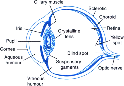
Accommodation: Accommodation is the ability or property of the eyelens due to which it can change its curvature or focal length so that images of objects at various distances can be formed on the same retina. The focal length of the eyelens is automatically changed with the help of ciliary muscles as follows:
(i) Viewing far off objects: When the ciliary muscles are completely relaxed, the eyelens is thin and its focal length is maximum (equal to distance between eyelens and retina). The rays coming from the distant object are parallel to each other and they are focussed at the retina as shown in Fig.(a).

Accommodation: Accommodation is the ability or property of the eyelen
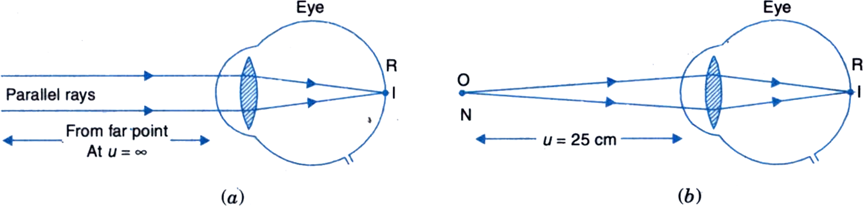
Fig. Accommodation of eyelens (a) Focussing parallel rays from infinity i.e., far point (b) Focussing rays from near point N.

(ii) Viewing nearby objects: When we look at a nearby object, the ciliary muscles contract, the eyelens bulges out and becomes thick and its focal length is reduced. This focusses the light from the nearby object on the retina, as shown in Fig. (b).

2312 Views

Advertisement

Describe the main parts of human eye. Explain its focussing action.


Human eye: It is the most valuable and sensitive sense organ. It is a remarkable optical instrument.

Human eye: It is the most valuable and sensitive sense organ. It is a
Fig. Structure of the human eye
Structure of the eye: As shown in Fig, 
the main parts of the human eye are as follows:
(i) Sclerotic: The eyeball is nearly spherical in shape with a diameter of about 2.3 cm. It has a tough and opaque white covering, called sclerotic which protects and holds the eye.
(ii) Cornea: Light enters the eye through a thin membrane called cornea which covers the transparent bulge on the front portion of the eyeball.
(iii) Choroid: It is a black membrane below the sclerotic. It absorbs stray light and avoids any blurring of image due to multiple reflections in the eyeball.
(iv) Iris: Behind the cornea, there is an opaque circular diaphragm called iris. The colour of the iris determines the colour of the eyes of a person. The iris has a central hole called the pupil. Due to its muscular action, the iris controls the size of the pupil and hence regulates the amount of light entering the eye. In bright light, the pupil becomes small. In dim light, the pupil opens up completely through the relaxation of the iris.
(v) Eyelens: It is a double convex lens situated behind the iris. It is composed of a fibrous, jelly like material. The lens is held in position by suspensory ligaments and connected to the sclerotic by the ciliary muscles. By contracting or relaxing, the ciliary muscles can change the shape or curvature of the eyelens and hence change its focal length. This ability of the eyelens to change its focal length is called accommodation. This enables the eyelens to focus the images of objects at different distances on the retina of the eye.
(vi) Retina: It is a delicate inner membrane on the backwall of the eyeball. It contains light sensitive cells called rods and cones. Rods are sensitive to intensity of light while cones are sensitive to colours. These cells change light energy into electrical signals which send message to the brain via the optic nerves.
(vii) Blind spot and yellow spot: In the region where the optic nerve enters the eyeball, there are no rods and cones. This region is totally insensitive to light and is called blind spot. Yellow spot has maximum concentration of light sensitive cells. It is situated in the centre of the retina.
(viii) Aqueous humour and vitreous humour: Aqueous humour is a salty fluid (n = 1.337) that fills the space between the cornea and the eyelens. Vitreous humour is a jelly like fluid (n = 1.437) that fills the space between the retina and the eyelens.
Focussing action of the eye: The transparent structures like cornea, aqueous humour, eyelens and vitreous humous together constitute a single converging lens. As the rays from an object enter the eye, they suffer refractions on passing successively through these structures and get converged. A real and inverted image is formed on the retina. The light sensitive cells of retina get activated and generate electrical signals that are sent to the brain through the optic nerves. Our brain translates the inverted image into an erect image.


3117 Views

Advertisement

Define the following terms and give their values for a normal eye:
(i) Range of normal vision.
(ii) Least distance of distinct vision.
(iii) Near point of the eye.
(iv) Far point of the eye.
(v) Power of accommodation.


(i) Range of normal vision: Due to accommodation property of the lens, a normal eye can clearly see the objects situated anywhere between infinity and 25 cm from it. At distance less than 25 cm, the ciliary muscles cannot bulge the eyelens any more, the object cannot be focussed on the retina and it appears blurred to the eye, as shown in Fig. The distance between infinity and 25 cm point is called the range of normal vision.

(i) Range of normal vision: Due to accommodation property of the lens
Fig. Object O within 25 cm from the eye is not focussed on retina and seen blurred
(ii) Least distance of distinct vision: The minimum distance from the eye, at which the eye can see the objects clearly and distinctly without any strain is called the least distance of distinct vision. It is denoted by the letter D. For a normal eye, its value is 25 cm.
(iii) Near point: The nearest point from the eye, at which an object can be seen clearly by the eye is called its near point. The near point of a normal eye is at a distance of 25 cm.
(iv) Far point: The farthest point from the eye, at which an object can be seen clearly by the eye is called the far point of the eye. For a normal eye, the far point is at infinity.
(v) Power of accommodation: The power of accommodation of the eye is the maximum variation of its power for focussing on near and far (distant) objects. For a normal eye, the power of accommodation is about four dioptres.


4023 Views

Give some points of similarity and dissimilarity between a camera and the human eye.

Points of similarity:

Camera

Human eye


1. Image is formed by a convex lens made of glass.

2. A real and inverted image is formed on the photographic film.

3. Diaphragm controls the amount of light entering the camera.

4. Time of exposure is controlled by a shutter.

1. Image is formed by the eyelens (a convex lens) made of fibrous, jelly like material.

2. A real and inverted image is formed on the retina.

3. Pupil in the iris controls the amount of light entering the eye.

4. Time of exposure is controlled by the eyelids.

Points of dissimilarity:

Camera

Human eye

1. Focal length of camera lens is fixed.

2. Focussing is done by changing the distance between the camera lens and the photographic film.

3. Photographic film retains the image permanently.

4. A photograph has to be changed for getting next image.

5. The angular region covered is about 60°.

1. Focal length of eyelens can be changed with the help of ciliary muscles.

2. Focussing is done by changing the shape of the eyelens by the action of ciliary muscles.

3.The retina of the eye retains the impression of an image for about (1/16)th of a second.

4. The same retina can be used for viewing an unlimited number of images.

5. The angular region covered is about 150°.

 
1890 Views

(a) What is myopia? State two reasons due to which this effect is caused.
(b) Draw a diagram to show the formation of image of a distant object by a myopic eye. How can such an eye defect be remedied?
Or
State two main causes of a person developing near sightedness. With the help of a ray diagram, suggest how he can be helped to overcome this disability.


Myopia or short-sightedness: It is a vision defect in which a person can see nearby objects clearly but cannot see the distant objects clearly beyond a certain point. This defect is common among children.
Cause of myopia: This defect arises due to either of the following two reasons:
(i) The eyeball gets elongated along its axis so that the distance between the eyelens and the retina becomes larger.
(ii) The focal length of the eyelens becomes too short due to the excessive curvature of cornea.

Myopia or short-sightedness: It is a vision defect in which a person
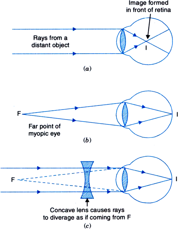
Fig. Myopic and its correction

As a result of the above causes, the parallel rays coming from a distant object do not meet at the retina but at a point in front of the retina, as shown in Fig. (a) and the distant object is not seen clearly. The object has to be moved closer to the eye to a point F to focus it on the retina, as shown in Fig. (b). Thus, the far point of a myopic eye is not at infinity but only a few metres from the eye.
Correction of Myopia: A myopic eye is corrected by using a concave lens of focal length equal to the distance of the far point F from the eye. This lens diverges the parallel rays from distant object as if they are coming from the far point F. Finally, the eyelens forms a clear image at the retina.


2826 Views

Advertisement