Describe the main parts of human eye. Explain its focussing action.
Human eye: It is the most valuable and sensitive sense organ. It is a remarkable optical instrument.
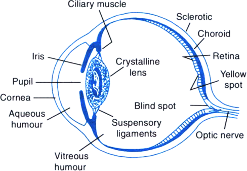
Fig. Structure of the human eye
Structure of the eye: As shown in Fig, the main parts of the human eye are as follows:
(i) Sclerotic: The eyeball is nearly spherical in shape with a diameter of about 2.3 cm. It has a tough and opaque white covering, called sclerotic which protects and holds the eye.
(ii) Cornea: Light enters the eye through a thin membrane called cornea which covers the transparent bulge on the front portion of the eyeball.
(iii) Choroid: It is a black membrane below the sclerotic. It absorbs stray light and avoids any blurring of image due to multiple reflections in the eyeball.
(iv) Iris: Behind the cornea, there is an opaque circular diaphragm called iris. The colour of the iris determines the colour of the eyes of a person. The iris has a central hole called the pupil. Due to its muscular action, the iris controls the size of the pupil and hence regulates the amount of light entering the eye. In bright light, the pupil becomes small. In dim light, the pupil opens up completely through the relaxation of the iris.
(v) Eyelens: It is a double convex lens situated behind the iris. It is composed of a fibrous, jelly like material. The lens is held in position by suspensory ligaments and connected to the sclerotic by the ciliary muscles. By contracting or relaxing, the ciliary muscles can change the shape or curvature of the eyelens and hence change its focal length. This ability of the eyelens to change its focal length is called accommodation. This enables the eyelens to focus the images of objects at different distances on the retina of the eye.
(vi) Retina: It is a delicate inner membrane on the backwall of the eyeball. It contains light sensitive cells called rods and cones. Rods are sensitive to intensity of light while cones are sensitive to colours. These cells change light energy into electrical signals which send message to the brain via the optic nerves.
(vii) Blind spot and yellow spot: In the region where the optic nerve enters the eyeball, there are no rods and cones. This region is totally insensitive to light and is called blind spot. Yellow spot has maximum concentration of light sensitive cells. It is situated in the centre of the retina.
(viii) Aqueous humour and vitreous humour: Aqueous humour is a salty fluid (n = 1.337) that fills the space between the cornea and the eyelens. Vitreous humour is a jelly like fluid (n = 1.437) that fills the space between the retina and the eyelens.
Focussing action of the eye: The transparent structures like cornea, aqueous humour, eyelens and vitreous humous together constitute a single converging lens. As the rays from an object enter the eye, they suffer refractions on passing successively through these structures and get converged. A real and inverted image is formed on the retina. The light sensitive cells of retina get activated and generate electrical signals that are sent to the brain through the optic nerves. Our brain translates the inverted image into an erect image.
3117 Views
