 Short Answer Type
 Long Answer TypeShort Answer TypeLong Answer Type
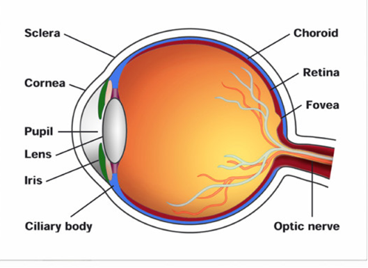
Eye Structure:
Eyes are spherical structures that consist of three layers.
(a) The outer layer is composed of sclera and cornea.
(i) Sclera - It is an opaque tissue that is usually known as white of the eye. It is composed of a dense connective tissue.
(ii) Cornea - It is a transparent anterior portion of the eye. It lacks blood vessels and is nourished by lymph from the nearby area. It is slightly bulged. It helps in focusing light rays.
(b) The middle layer of the eye is vascular in nature and contains choroid, ciliary body, and iris.
(i) Choroid lies next to the sclera and contains numerous blood vessels that provide nutrients and oxygen to the retina and other tissues.
(ii) Ciliary body: The choroid layer is thin over posterior region and is thick in the anterior portion to form ciliary body. It contains blood vessels, ciliary muscles, and ciliary processes.
(iii) Iris: It is the visible thin coloured portion present at the junction of sclera and cornea.
Lens - The eye contains a transparent, biconvex, and elastic structure just behind the iris. It is known as lens. The lens is held in position by suspensory ligaments attached to the ciliary body. The lens divides the eye ball into two chambers – an anterior aqueous and posterior vitreous chamber.
(c) The innermost nervous coat is the Retina. It contains three layers of cells – inner ganglion cells, middle bipolar cells, and outermost photoreceptor cells.
The receptor cells present in the retina are of two types – rod cells and cone cells.
(a) Rod cells –The rods contain the rhodopsin pigment (visual purple) that is highly sensitive to dim light. It is responsible for twilight vision.
(b) Cone cells –The cones contain the iodopsin pigment (visual violet) and are highly sensitive to high-intensity light. They are responsible for daylight and colour visions.
The innermost ganglionic cells give rise to optic nerve fibre that forms optic nerve in each eye and is connected to the brain.


 Switch


